Perioperative management - Total gastrectomy, robotically assisted with D2 lymphadenectomy

  1. Indications

    506_Anatomie_Lymphe besch2 Kopie.jpg

    Primary Gastric Carcinoma

    In a curative therapeutic approach and local resectability, there is generally an indication for surgery, provided there are no significant risk factors (e.g., pre-existing conditions or the general condition of the patient; see also contraindications).

    For locally advanced tumors of stages II and III (cT1/cT2N+ and cT3/resectable cT4Nx), pre- or perioperative chemotherapy should be performed.

    To achieve tumor-free resection margins (R0), a proximal safety margin of 5 cm (for the intestinal type according to Lauren) or 8 cm (for the diffuse type according to Lauren) is generally required, except for mucosal carcinomas (T1a N0 M0). The extent of resection (total versus subtotal gastrectomy) is determined by the tumor location and spread, as well as the histological type, which requires an appropriate safety margin.

    Early Gastric Carcinoma

    An early gastric carcinoma is defined as a tumor that, regardless of lymph node status, surface area, and distant metastasis, is confined to the mucosa and submucosa of the stomach wall. The muscularis propria of the stomach is tumor-free by definition.

    In early carcinomas, the frequency of lymph node metastasis varies. Tumors that have already infiltrated the submucosa are not eligible for endoscopic therapy, as there is a 4-20% probability of lymph node metastasis.

    About 5% of patients show an early gastric carcinoma of the mucosal type (pT1m), where curative therapy through endoscopic resection is possible, as the probability of lymph node metastasis is extremely low. Affected patients have an excellent prognosis with a five-year survival rate of over 90%.

    Indications for Endoscopic Resection

    Superficial gastric carcinomas confined to the mucosa (about 5% of patients) can be treated with endoscopic resection considering the following criteria:

    • Lesions < 2 cm in size for elevated types
    • Lesions up to 1 cm in size for flat types
    • Histological differentiation grade: well or moderately (G1/G2)
    • No macroscopic ulceration
    • Invasion confined to the mucosa

    Special Situations:

    Gastric Stump Carcinoma (Recurrent Tumors after Subtotal Gastrectomy):
    Completion gastrectomy resulting in total gastrectomy.

    Oligometastasis:
    As part of a multimodal approach, within studies and when technically feasible, all metastatic sites can be removed. Metastases discovered intraoperatively can, similar to the esophageal guideline, be resected if they are R0-resectable.

    Peritoneal Metastases:

    Requirements:

    • PCI < 12
    • Complete resection of all macroscopically visible tumor parts possible

    Procedure:

    • Neoadjuvant systemic therapy
    • Oncological resection (gastrectomy/subtotal gastric resection) with D2 lymphadenectomy and tumor-free resection margins
    • Complete resection of peritoneal metastases (cytoreductive surgery)
    • Hyperthermic intraperitoneal chemotherapy

    In a palliative therapeutic approach, gastrectomy may be indicated in rare cases (e.g., in bleeding, perforation, stenosis).

    Lymphadenectomy

    The lymph node dissection of compartments I and II is referred to as D2-LAD and represents the standard lymphadenectomy for gastric carcinoma. It is considered the gold standard.

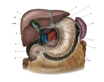
    Based on the Japanese classification (numbering), the lymph nodes are grouped into several compartments. The D1 lymph nodes include stations 1 to 6, the D2 compartment additionally includes stations 7 to 11.

    The lymph nodes of station 12 are resected in the context of oncological resection for distally located carcinomas on the lesser curvature side. Lymph nodes at the main bile duct (station 12b, considered M1) are often not resected to avoid injury/devitalization of the bile duct.

    In the D3 compartment, groups 12 to 15 are located, which by definition are not regional lymph node stations of the stomach. As they are prognostically considered distant metastases, they are classified as M1 LYM if involved.

    To achieve a classification of pN0, the UICC requires a minimum of 16 regional lymph nodes to be examined. The German S3 guideline specifies an intended lymph node count of ≥ 25.

  2. Contraindications

    Patient-specific:

    • Severely reduced general condition (e.g., due to tumor cachexia)
    • ECOG status ≥ 2
    • Relevant comorbidities with inability to undergo surgery or anesthesia, e.g.:
      • Liver cirrhosis Child B and C
      • Poor cardiac and pulmonary function (NYHA status and GOLD status)
      • High-grade carotid stenoses before therapy
      • Adhesive abdomen in relation to a minimally invasive approach

    Tumor-related unresectability:

    • Unresectable tumor with proximal infiltration of the superior mesenteric artery or the celiac trunk
    • Any form of metastasis, unless the metastasis can be completely treated by R0 resection (e.g., locally limited peritoneal carcinomatosis, solitary liver metastasis, solitary distant lymph node metastasis).
    • Complete portal vein thrombosis or other conditions with pronounced venous collateral circulation (e.g., liver cirrhosis).
    • In case of passage obstruction or endoscopically uncontrollable tumor bleeding, gastrectomy may be indicated as a palliative procedure.
  3. Preoperative Diagnostics

    Mandatory:

    • Laboratory tests with so-called tumor markers (CA 72-4, CA 19-9, CEA)
    • Esophagogastroduodenoscopy with step biopsies (at least 8 biopsies)
    • Abdominal ultrasound: First imaging procedure for assessing liver metastases.
    • Endosonography for assessing the T-stage, particularly for evaluating early gastric cancer (mucosal/submucosal type). Identification of risk constellations uT3/4 N+ or detection of perigastric ascites. Assessment of lymph nodes in category N1–2 is possible, though with limited sensitivity and specificity.
    • Abdomen/pelvis CT (with i.v. contrast): Local situation, lymph nodes, liver metastases
    • Thoracic CT to rule out pulmonary metastases
    • MRI: For patients who cannot undergo CT.
    • PET-CT is not routinely recommended for staging gastric carcinomas.

    Additionally for T3/T4:

    Staging Laparoscopy:

    Goal:
    To clarify peritoneal metastasis. Since peritoneal metastasis cannot yet be reliably excluded by imaging, diagnostic laparoscopy is generally recommended for patients with locally advanced gastric cancer (T3–T4) before starting neoadjuvant chemotherapy. In up to 40% of cases, peritoneal metastasis is detected.

    Advantages of Laparoscopy:

    • In addition to the possibility of histological confirmation of peritoneal metastases, diagnostic laparoscopy helps minimize unnecessary open exploratory attempts.
    • It allows the detection of previously unknown small subcapsular liver metastases.
    • In combination with intraoperative ultrasound (IOUS) and biopsy, metastases can be identified even more precisely.

    Peritoneal lavage with cytology:
    A peritoneal lavage with cytology can be performed additionally. The probability of metachronous peritoneal metastasis is about 80% if a positive lavage cytology is present. Currently, studies are ongoing to identify patients who could benefit from additional therapeutic measures (e.g., HIPEC) despite a macroscopically negative finding.

    Trocars and access route:
    To enable complete parietal peritonectomy in the event of peritoneal metastasis, the trocars should always be placed in the linea alba during diagnostic laparoscopy. Usually, two trocars are sufficient:

    • A 10mm trocar for the optics
    • A 5mm trocar for the biopsy forceps

    Trocar metastases:
    Trocar metastases that occur later can also be easily removed during a median laparotomy.

  4. Special Preparation

    Neoadjuvant chemotherapy as an essential part of the treatment concept for gastric carcinoma

    For the diagnosis of gastric carcinoma stages II and III (cT1/cT2N+ and cT3/resectable cT4Nx), a perioperative chemotherapy should be performed according to the current guideline. This is divided into two phases:

    Preoperative chemotherapy (Neoadjuvant therapy):

    • Start of chemotherapy before surgery.
    • The goal is to shrink the tumor to improve resectability and increase the likelihood of complete tumor removal.

    Postoperative chemotherapy (Adjuvant therapy):

    • Continuation of chemotherapy after surgery.
    • The aim is to eliminate microscopic residual tumors or possible microscopic metastases, thereby reducing the risk of recurrence.

    Through this perioperative approach, the R0 resection rate (tumor-free) can be increased and the systemic recurrence rate reduced. This leads to an improved overall prognosis for the patients.

    FLOT regimen:

    • FLOT stands for a combination of four chemotherapeutic agents: 5-Fluorouracil, Leucovorin, Oxaliplatin, and Docetaxel.
    • This regimen has been established as the standard for perioperative chemotherapy both in Germany and internationally.
    • The prognostic value of this treatment approach is still under discussion. There are considerations whether it is an overtreatment or if it actually represents a meaningful concept.
      (Gockel I, Lordick F. Neoadjuvant chemotherapy in gastric carcinoma. Chirurg. 2020 May;91(5):384-390.)

    Evaluation of response to therapy:

    After completion of neoadjuvant therapy, it is recommended to assess the response to treatment using CT and endoscopy to evaluate the effectiveness of the therapy and plan further procedures.

    In planned preoperative chemotherapy:

    Port implantation:

    • Before starting chemotherapy, a port is implanted to ensure access to the chemotherapeutic agents.

    Laparoscopic exploration to clarify peritoneal carcinomatosis:

    • This serves for the early detection of peritoneal metastasis, which may not be reliably detectable with imaging techniques.

    Possibly stent placement in case of dysphagia:

    • If the tumor causes dysphagia (swallowing difficulties), a stent can be placed to support food intake.

    In planned resection:

    Preoperative nutrition:

    • Possibly initiating preoperative nutritional therapy in patients who have significantly lost weight preoperatively.
    • A nutritional concept should be developed early, preferably at the beginning of multimodal therapy.
    • Even patients without signs of malnutrition should take balanced oral nutritional supplements for 5–7 days in addition to their normal diet to optimize their food intake.

    The day before the surgery:

    • Current laboratory tests: To determine relevant values such as blood type and order erythrocyte concentrates.
    • Thrombosis prophylaxis: Thrombosis prophylaxis with low molecular weight heparin (LMWH) and anti-thrombosis stockings is performed according to the guideline for thrombosis prophylaxis.
  5. Informed consent

    General Surgical Risks:

    • Thrombosis, Pulmonary Embolism
    • Wound Healing Disorder
    • Injury to Internal Organs (e.g., intestines, liver, spleen)
    • Follow-up Interventions
    • Bleeding/Rebleeding
    • Pneumonia
    • Urinary Tract Infection
    • Heparin Intolerance, HIT (Heparin-induced Thrombocytopenia)

    Specific Surgical Risks:

    • Conversion to an Open Procedure
    • Mortality between 2 and 10%
    • Final Decision on Resection Procedure only intraoperatively
    • Anastomotic Insufficiency
    • Duodenal Stump Insufficiency
    • Endoluminal/Intra-abdominal Bleeding
    • Reduced Blood Supply to the Substitute Stomach
    • Intra-abdominal Abscess, Peritonitis
    • Pancreatitis or Pancreatic Fistulas
    • Wound Healing Disorder
    • Incisional Hernia

    Potentially Necessary Extension of the Procedure:

    • Cholecystectomy
    • Simultaneous Resection of Liver Metastases
    • Splenectomy

    Indications of:

    • Temporary Weight Loss
    • Changes in Eating Habits
    • Drains, Nasogastric Tube, Urinary Diversion
    • Possible Blood Transfusion
Anesthesia

Intubation anesthesiaNasogastric tubeCVC (Central Venous Catheter)Epidural catheterIndwelling cathe

Activate now and continue learning straight away.

Single Access

Activation of this course for 3 days.

€7.99 inclusive VAT

Most popular offer

webop - Savings Flex

Combine our learning modules flexibly and save up to 50%.

from €3.70 / module

€44.50 / yearly payment

price overview

Robotik

Unlock all courses in this module.

€7.42 / month

€89.00 / yearly payment