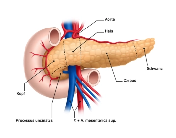
- The pancreas, composed of lobules, has a reddish-gray color, is 14 – 18 cm long, and weighs 65 – 80 grams. It is located at the level of the 1st and 2nd lumbar vertebrae and extends wedge-shaped from the epigastric region into the left hypochondriac region.
- The pancreas is enclosed by capsule-like connective or fatty tissue and is divided into four sections: head, neck, body, and tail. While a somewhat firmer connective tissue plate is located in the posterior head area, the gland is otherwise predominantly loosely connected dorsally with the connective tissue. As a retroperitoneal organ, the gland is covered with peritoneum on its anterior surface.
- The widest part of the gland is the head of the pancreas, which, located to the right of the spine, fits into the loop formed by the duodenum. Both the anterior and posterior surfaces of the duodenum can be overlaid by glandular tissue to varying degrees here. The head encompasses with its caudal part (uncinate process) from behind the superior mesenteric vein and artery. The groove located in the uncinate process and the remaining part of the pancreatic head is referred to as the pancreatic notch.
- The part of the pancreas located at the level of the 1st lumbar vertebral body represents the transition area from the head to the body, with a width of about 2 cm, and lies over the superior mesenteric vessels. From a surgical perspective, this section is also referred to as the neck of the pancreas.
- The elongated body of the pancreas runs obliquely upward over the 1st and 2nd lumbar vertebrae, bulges ventrally into the omental bursa, and arches towards the splenic hilum, with the transition into the tail occurring without precise anatomical demarcation. Dorsal to the body, alongside the spine, are the aorta, inferior vena cava, and superior mesenteric artery and vein.
- The tail of the pancreas forms the pointed continuation of the glandular body and extends to or into the splenorenal ligament.
- The pancreas can be arranged in various morphological variants, including oblique, S-shaped, transverse, and L-shaped. A horseshoe shape and an inverted V-shape have also been described. The transitions between the morphological variants are fluid.
-
Relevant macroscopic anatomy
![513_Pankreasanatomie.jpeg]()
-
Spatial relationships to other organs and conduits
![Spatial relationships to other organs and conduits]()
- Developmentally, the organ is in close proximity to the upper abdominal organs and vessels.
- Topographically, the pancreas has multiple relationships with adjacent organs and retroperitoneal pathways:
- ventrally, the omental bursa and the posterior surface of the stomach
- to the right, there is a close relationship between the head and the duodenal C
- to the left, the tail of the pancreas extends to the splenic hilum
- the posterior wall of the pancreas touches at the level of the head the portal vein, the superior mesenteric artery and vein as well as the common bile duct, at the level of the body the splenic artery and vein, the inferior mesenteric vein, the inferior vena cava and the abdominal aorta, at the level of the tail the left kidney
Note: Surgically relevant are the following anatomical details
- The inferior mesenteric vein runs under the lower border of the pancreas.
- There is a close relationship between the spleen and stomach in the area of the short gastric arteries and veins,
-
Pancreatic duct system
The approximately 2 mm thick Ductus pancreaticus (Wirsungianus) traverses the organ in its longitudinal extension near the posterior surface and receives numerous short ductal branches that enter it perpendicularly along its course. In about 77% of cases, the duct joins with the common bile duct at the Papilla duodeni major (Papilla Vateri) in the posterior wall area of the descending part of the duodenum; in the remaining cases, the openings of both ducts are located close to each other. The Ductus pancreaticus accessorius, an accessory duct, is often only rudimentarily developed or completely absent. If present, it opens at the Papilla duodeni minor (Papilla Santorini).

Vascular supply
The vascular supply of the pancreas is ensured by numerous vessels.Head: The arterial supply of the
The vascular supply of the pancreas is ensured by numerous vessels.Head: The arterial supply of the
Activate now and continue learning straight away.
Single Access
Activation of this course for 3 days.
US$9.30
inclusive VAT
Most popular offer
webop - Savings Flex
Combine our learning modules flexibly and save up to 50%.
from US$4.33 / module
US$52.04/ yearly payment
robotics
Unlock all courses in this module.
US$8.67
/ month
US$104.10 / yearly payment
Webop is committed to education. That's why we offer all our content at a fair student rate.