Anatomy - Toupet Fundoplicatio for GERD, Robot-Assisted

  1. Surgical Anatomy of the Stomach

    505_Anatomie_Magen_01.jpeg

    The stomach is a muscular hollow organ located between the esophagus and duodenum, situated in the left and central upper abdomen directly beneath the diaphragm. When moderately filled, it is 25 – 30 cm long and has a storage capacity of 1.5 liters, which can extend up to 2.5 liters in extreme cases. The position, size, and shape of the stomach vary greatly depending on age, filling state, and body position, with significant interindividual differences.

    The stomach is divided into the following sections:

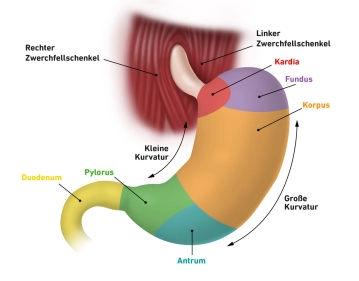
    • Cardia (Gastric Entrance, Upper Gastric Orifice, Ostium cardiacum):
      The cardia is a 1–2 cm region where the esophagus transitions into the stomach. The sharp junction between the esophageal and gastric mucosa, often visible endoscopically, marks this area
       
    • Fundus gastricus (Gastric Fundus):
      Located above the gastric entrance, the fundus arches upward, also known as the gastric dome or Fornix gastricus. It is usually filled with air swallowed involuntarily during eating. In an upright position, the fundus forms the highest point of the stomach, and the collected air appears as a “gastric bubble” in radiographic images. It is separated from the cardia by a distinct fold, the Incisura cardialis
       
    • Corpus gastricum (Gastric Body):
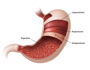
      The main part of the stomach, the corpus, features deep longitudinal mucosal folds (Plicae gastricae), extending from the cardia to the pylorus. These folds are also referred to as the “gastric road”
       
    •  Pylorus (Pars pylorica, Gastric Pylorus):
      This section begins with the expanded Antrum pyloricum, followed by the pyloric canal (Canalis pyloricus), and ends with the pylorus itself. The pylorus contains the pyloric sphincter (M. sphincter pylori), a thick circular muscle that closes the lower gastric orifice (Ostium pyloricum), allowing periodic passage of chyme into the duodenum

    The stomach is intraperitoneal, covered by serosa except for the dorsal cardia. The embryonic mesogastria rotate from a sagittal to a frontal position during development:

    • The Omentum minus extends from the lesser curvature to the liver hilum
    • The Omentum majus extends from the greater curvature to the transverse colon, spleen, and diaphragm

    The stomach is located intraperitoneally, which gives it a serosal covering, except for the posterior portion of the cardia, which lacks serosa. During embryonic development, the mesogastria undergo a rotation from their original sagittal orientation to a frontal position. The omentum minus extends from the lesser curvature to the porta hepatis, while the omentum majus spreads out from the greater curvature to the transverse colon, spleen, and diaphragm.

    The stomach is anchored and stabilized within the abdominal cavity by ligaments that extend to the liver and spleen. Its convex side forms the greater curvature (greater gastric curvature/curvatura major), while its concave side creates the lesser curvature (lesser gastric curvature/curvatura minor). The anterior wall is referred to as the paries anterior, and the posterior wall as the paries posterior. The greater curvature gives rise to the omentum majus, and the omentum minus stretches between the left liver lobe and the lesser curvature.

  2. Layers and Structure of the Gastric Wall

    505_Anatomie_Magen_02.jpeg

    The gastric wall has a characteristic layered structure when viewed microscopically, progressing from the innermost to the outermost layers:

    Tunica mucosa (Mucosal Layer):
    This layer lines the stomach’s interior and consists of three sublayers:

    • Lamina epithelialis mucosae: Produces a thick, neutral mucus that protects the gastric mucosa from mechanical, thermal, and enzymatic damage
    • Lamina propria mucosae: Contains the gastric glands (Glandulae gastricae)
    • Lamina muscularis mucosae: A thin muscular layer capable of altering the mucosal relief

    Tela submucosa (Submucosal Layer):
    This loose connective tissue layer provides a sliding surface and contains:

    • A dense network of blood and lymphatic vessels
    • The Plexus submucosus (Meissner’s plexus): Regulates gastric secretion, functioning autonomously with modulation by the autonomic nervous system

    Tunica muscularis (Muscular Layer):
    A robust layer subdivided into:

    • Fibrae obliquae: Inner oblique muscle fibers
    • Stratum circulare: Middle circular muscle layer
    • Stratum longitudinale: Outer longitudinal muscle layer

    These layers enable gastric peristalsis, mixing chyme with gastric juice. Between the circular and longitudinal layers lies the Plexus myentericus (Auerbach’s plexus), which autonomously regulates muscle function while receiving input from the autonomic nervous system.

    Tela subserosa (Subserosal Layer):
    A connective tissue layer providing additional structural support.
     

    Tunica serosa (Serosal Layer):
    The outermost layer consists of:

    • Lamina propria serosae: Contains blood vessels, lymphatics, nerves, and immune cells (e.g., Macula lactea or milk spots)
    • Lamina epithelialis serosae: A single-layered squamous epithelium providing a slippery surface to reduce friction with adjacent organs

    Gastric Glands (Glandulae gastricae):
    Gastric glands are located in the Lamina propria mucosae and are particularly abundant in the fundus and corpus. Around 100 glands per mm² are present in the mucosa, composed of different cell types:

    • Mucous cells: Produce neutral mucus, identical to the epithelial cells’ secretion
    • Neck cells: Located near the gland’s surface; secrete alkaline mucus rich in bicarbonate ions to regulate pH and protect against self-digestion. These cells are abundant in the cardia and fundus
    • Chief cells: Produce pepsinogen, an inactive precursor of pepsin, which activates upon contact with hydrochloric acid (HCl) at the gland’s surface. These cells are concentrated in the gastric corpus
    • Parietal cells (Belegzellen): Found primarily in the corpus; they produce:
      • Hydrogen ions (H⁺) for HCl synthesis, resulting in a gastric pH of 0.9 – 1.5
    • Intrinsic factor: essential for forming a complex with dietary vitamin B12 to enable absorption through the intestinal wall. This factor is critical for erythropoiesis; its absence after gastrectomy may lead to anemia
    • G-cells: Predominantly located in the antrum, these cells secrete gastrin, stimulating HCl production in parietal cells

    This intricate structure enables the stomach to perform its functions of digestion, mechanical mixing, and protection from its acidic environment.

  3. Function

    The stomach acts as a reservoir for ingested food, storing and mixing it. It produces acidic gastric juice (mucus and HCl) and enzymes that begin the digestion of some food components. The resulting chyme is then gradually released into the duodenum through the pylorus in a regulated manner.

    The stomach can store food for several hours, allowing for fewer, larger meals to meet daily nutritional needs.

  4. Arterial Supply

    505_Anatomie_Arterien besch.jpg

    The arterial blood supply to the stomach is derived from branches of the unpaired celiac trunk. These vessels run along the gastric curvatures as vascular arcades, forming numerous anastomoses:

    • Arteria gastrica dextra: From the proper hepatic artery, supplying the lower portion of the lesser curvature
    • Arteria gastrica sinistra: Supplying the upper portion of the lesser curvature
    • Arteriae gastricae breves: From the splenic artery, supplying the fundus
    • Arteria gastroepiploica (omentalis) dextra: From the gastroduodenal artery, supplying the lower (right) portion of the greater curvature
    • Arteria gastroepiploica (omentalis) sinistra: From the splenic artery, supplying the left portion of the greater curvature
    • Arteria gastrica posterior: From the splenic artery, supplying the posterior gastric wall

    The stomach is nourished by two major vascular arcades:

    • Along the lesser curvature (formed by the left and right gastric arteries)
    • Along the greater curvature (formed by the left and right gastroepiploic arteries)
Venous and neural Supply

Venous Supply The venous drainage of the stomach parallels its arterial supply, with four main vein

Activate now and continue learning straight away.

Single Access

Activation of this course for 3 days.

€7.99 inclusive VAT

Most popular offer

webop - Savings Flex

Combine our learning modules flexibly and save up to 50%.

from €3.29 / module

€39.50 / yearly payment

price overview

Robotik

Unlock all courses in this module.

€7.42 / month

€89.00 / yearly payment