Anatomy - Inguinal hernia repair, Lichtenstein

  1. Surgically relevant anatomy

    Surgically relevant anatomy

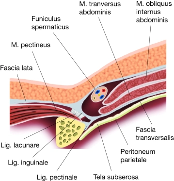
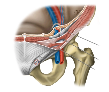
    The inguinal region (transition between the anterior abdominal wall and the lower extremity) has several weak points through which a hernial sac with or without contents can protrude through the abdominal wall (women are more prone to femoral hernias, men to inguinal hernias). Below the inguinal ligament, the inguinal canal is divided into two compartments by a division of the inguinal ligament (arcus iliopectineus): the lacuna vasorum and the lacuna musculorum.

    Lacuna vasorum

    • Located next to the pubic bone, it serves as the passageway for the external iliac artery and vein (→ femoral artery and vein, arrangement: artery lateral to the vein). The femoral branch of the genitofemoral nerve passes through it laterally. Caudally medial, the deep inguinal lymph nodes (Rosenmüller) are found here. The lacuna vasorum represents the internal hernial orifice for femoral hernias (through the femoral septum next to the femoral vein).

    Lacuna musculorum

    • Located lateral to the lacuna vasorum, it serves as the passageway for the psoas major muscle and iliacus muscle (together = iliopsoas muscle) as well as for the femoral nerve and lateral femoral cutaneous nerve (cranially).
  2. Anterior abdominal wall and inguinal canal

    Anterior abdominal wall and inguinal canal

    The inguinal canal forms during the development of the gonads in the male embryo through the descent of the testes, which are drawn into the scrotum by the gubernaculum testis, as a tubular structure. The peritoneum carried along during this descent remains as an outpouching in the inguinal canal (Processus vaginalis testis) and extends to the epididymis. The layers of the abdominal wall thus become comparable coverings in this pouch:

    • Fascia transversalis → Internal spermatic fascia,
    • Internal oblique muscle → Cremaster muscle,
    • Fascia of the external oblique muscle → External spermatic fascia,
    • no covering by the transversus abdominis muscle, as it ends more cranially.

    Blood vessels (testicular artery and vein) as well as the vas deferens (ductus deferens) and nerves (ilioinguinal nerve from the lumbar plexus) are also drawn into the scrotum and form the spermatic cord. Through the obliteration of the processus vaginalis testis, the connection to the abdominal cavity closes, typically leaving only its entrance (vestige of the processus vaginalis testis). In the female embryo, the ovaries do not completely descend due to the pull of the gubernaculum, but remain beside the uterus. Only the round ligament of the uterus, as the former gubernaculum, persists in the inguinal canal. Inadequate obliteration of the processus vaginalis testis represents a weakness in the abdominal wall and the starting point for inguinal hernias.

    The inguinal canal runs approximately 4 cm long in a medio-caudal direction just above the inguinal ligament parallel to it and lies between the deep inguinal ring, as its cranial end, and the superficial inguinal ring, as its opening to the external abdominal wall.

    Deep inguinal ring

    • Midway between the symphysis and the anterior superior iliac spine in the inner abdominal wall, lateral to the inferior epigastric artery and vein (in the lateral umbilical fold)

    Superficial inguinal ring

    • Above the pubic tubercle in the fascia of the external oblique muscle, upper edge pointing cranially, lower edge formed by the inguinal ligament, sides = medial and lateral crus with intercrural fibers for stabilization.
  3. Walls of the inguinal canal

    Anterior Wall

    • Fascia of the external oblique muscle, laterally reinforced by fibers of the internal oblique muscle → inguinal ligament and → cremaster muscle

    Posterior Wall

    • Transversalis fascia, medially reinforced by the inguinal falx (= tendon of the transversus abdominis muscle and internal oblique muscle)

    Superior Wall

    • Transversus abdominis muscle and internal oblique muscle (fiber direction from the inguinal ligament to the medial inguinal falx)

    Inferior Wall

    • Medial inguinal ligament (=ligamentum reflexum) as well as a groove formed by the external oblique muscle for the spermatic cord.
  4. Contents of the inguinal canal

    In males, the inguinal canal contains the spermatic cord (Funiculus spermaticus) with the ductus deferens, the artery of the ductus deferens (from the inferior vesical artery) and the testicular artery (from the aorta), the venous pampiniform plexus, the cremasteric artery/vein, the genital branch of the genitofemoral nerve to the cremaster muscle, as well as sympathetic nerve fibers and lymphatic vessels, surrounded by the internal spermatic fascia, the cremasteric fascia, and the external spermatic fascia.

    In females, the inguinal canal contains the round ligament of the uterus, which extends from the uterus to the deep inguinal ring, through the inguinal canal to the superficial inguinal ring, and finally ends at the labia majora.

    Additionally, lymphatic vessels are present. The ilioinguinal nerve partially runs in the inguinal canal.

  5. Types of hernias

    Hernial openings for indirect inguinal hernias are the deep inguinal ring, and for direct inguinal hernias, the medial inguinal fossa (medial to the deep inguinal ring and the inferior epigastric artery/vein).

    Indirect Inguinal Hernias

    • more common, men > women, congenital (patent processus vaginalis) or acquired (also through the internal inguinal ring in the lateral inguinal fossa, mostly adults), the hernia sac runs lateral to the epigastric vessels and extends into the scrotum or labia majora

    Direct Inguinal Hernias

    • mostly acquired, in adults men > women, protrusion of the peritoneum and transversalis fascia in the medial inguinal fossa (= Hesselbach's triangle/inguinal triangle, medial to the epigastric artery/vein), medially bounded by the transversus abdominis muscle, caudally by the inguinal ligament, mostly exiting through the external inguinal ring → scrotum/labia majora.

    Femoral Hernias

    • predominantly acquired, in adults women > men, through the femoral canal in the medial thigh (medial to the vascular lacuna) along with the femoral artery/vein, femoral branch of the genitofemoral nerve, and lymphatic vessels.