Anatomy - Total laparoscopic gastrectomy with D2 lymphadenectomy

  1. Surgical anatomy of the stomach

    Surgical anatomy of the stomach

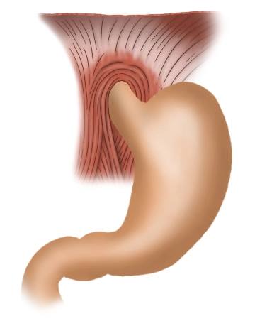
    In terms of function, the stomach mixes and stores food and is an expansion of the alimentary tract between the esophagus and the duodenum. This muscular hollow viscus produces acidic gastric juice (mucus and HCl) and enzymes, which predigest some elements of the ingested food, and portions the chyme into the duodenum.

    Usually, the stomach is located immediately inferior to the diaphragm in the left upper quadrant and epigastrium. Location, size and shape of the stomach vary from person to person and may differ substantially, depending on age, filling condition and body position. The moderately filled stomach has a mean length of 25- 30 cm and can hold 1.5 liters, in extreme cases up to 2,5 liters.

    Within the abdominal cavity the stomach is held in position and stabilized by ligaments inserting at the liver and spleen Its convex aspect forms the major curvature (curvatura major gastrica) and its concave aspect the lesser curvature (curvatura minor gastrica). Its anterior wall is termed paries anterior gastrica and its posterior aspect paries posterior gastrica.

    Since the stomach is an intraperitoneal viscus, it is covered by the gastric serosa (tunica serosa gastrica), and only the posterior aspect of the cardia is free of serosa. Stomach rotation shifts the embryonic mesogastrics from their former sagittal position to a frontal location. The lesser omentum originates at the lesser curvature and extends to the hepatic portal, while the greater omentum originates at the greater curvature and courses to the transverse colon, spleen and diaphragm.

    The stomach displays the following portions:

    Entrance of stomach / Cardia / Ostium cardiacum:
    The superior opening of the stomach, where the esophagus enters the stomach, is 1- 2 cm long. It is characterized by a marked transition from the mucosa of the esophagus to that of the stomach.

    Gastric fundus / fundus gastricus:
    Superior to the level of entrance of the esophagus the fundus arches cephalad, which then is called gastric fornix (fornix gastricus). Usually, the fundus is full of air which is swallowed automatically when ingesting food. In the erect position the fundus is the highest point of the stomach, and on abdominal films its trapped air is evident as the “gastric bubble”. A notch (incisura cardialis) clearly delimits the fundus from the entrance of the stomach.

    Body of the stomach / Corpus gastricum
    The main portion of the stomach is taken up by the gastric body. The deep mucosal folds (plicae gastricae) found here extend from the cardia to the pylorus and are also known as “magenstrasse”.

    Pylorus / Pars pylorica:
    This portion begins with the extended pyloric antrum, followed by the pyloric canal, and terminates at the actual pylorus. It is formed by the pyloric sphincter (m. sphincter pyloricus), a strong circular layer of muscle which closes off the inferior gastric orifice (ostium pyloricum). The pylorus closes off the gastric outlet and periodically lets some of the chyme pass into the adjacent duodenum.
  2. Layers and structure of the gastric wall

    wall Gastric

    Under the microscope the gastric wall displays a characteristic layered structure with the following sequence from the inside out:

      • The internal aspect of the gastric wall is lined by mucosa (tunica mucosa). The gastric mucosa is made up of three sublayers: The lamina epithelialis mucosae produces viscous neutral mucus which protects the gastric mucosa against mechanical, thermal and enzymatic injury. This is followed by the loose connective tissue coat of the lamina propria mucosae into which the gastric glands (glandulae gastricae) descend. The outermost layer of the mucosa is the small lamina muscularis mucosae which can change the relief of the mucosa.
      • The gastric mucosa is followed by a loose layer of connective tissue (tela submucosa gastrica), which houses not only a dense network of blood and lymph vessels but also a nerve plexus (plexus submucosus or Meissner plexus) which controls gastric secretion. Although this plexus is independent of the central nervous system (CNS), the latter may affect the former via the autonomic nervous system.
      • Next is the marked tunica muscularis with its three sublayers, each comprising muscle fibers coursing in different directions: The inner layer of small , oblique muscle fibers (fibrae obliquae), then a circular layer (stratum circulare) and finally the outermost longitudinal layer of muscle fibers (stratum longitudinale). These muscles effect the peristalsis of the stomach and ensure thorough mixing of the chyme with the gastric juice. Muscular function is controlled by a nerve plexus, the plexus myentericus or Auerbach plexus, in between the circular and longitudinal layers. Just like the plexus submucosus, this plexus is mostly autonomous but is also affected by the autonomic nervous system.
      • Next is another layer of loose connective tissue (tela subserosa gastrica).
      • The peritoneum (tunica serosa) covering the external aspect of the stomach is its final layer.

    Gastric glands The gastric glands (glandulae gastricae) located in the fundus and body of the stomach are part of the lamina propria mucosae. 1 mm2 of mucosal surface comprises up to 100 such glands. The ductal wall of the gland is lined with different types of cells:

      • Mucous cells: They produce the same neutral mucus as the epithelial cells.
      • Surface mucous cells: Foveolar cells are close to the surface of the gland and contain alkaline mucus, i.e., the pH of its hydrogen carbonate ions (HCO3–) is rather high. This property is rather important in controlling the gastric pH. The mucus lines the gastric mucosa and protects it against autodigestion by the aggressive hydrochloric acid (HCl) and enzymes as autodigesting proteins. This type of cells is mostly found in the cardia and fundus of the stomach.
      • Chief cells: These cells produce the inactive proenzyme pepsinogen which, once released, is activated by hydrochloric acid (HCl) to the active enzyme pepsin, the latter starting the digestion of the alimentary proteins. Since the initial contact of the enzyme with hydrochloric acid is at the surface of the gland, this ensures that the glands will not be autodigested by the enzyme. This type of cells is mostly found in the body of the stomach.
      • Parietal cells: Mostly found in the body of the stomach, these cells produce plenty of hydrogen ions (H+) needed in the production of hydrochloric acid (HCl). The latter has a rather low pH of 0.9- 1.5. In addition, the parietal cells also produce the so-called intrinsic factor and. Together with vitamin B12 from the ingested food this substance generates a complex in the small intestine which is able to pass through the intestinal wall. This vitamin plays a pivotal role in erythropoiesis (gastric resection may result in anemia).
      • G cells: Primarily found in the gastric antrum, these cells produce gastrin which increases HCl production in the parietal cells.
  3. Function

    The stomach acts as a reservoir for ingested food. Since it may store food for hours, it ensures that we can meet our daily nutritional requirements with a few major meals. Peristalsis thoroughly mixes the chyme with the gastric juice, the food is broken up chemically, predigested and then portioned into the duodenum.

Arterial and venous blood supply, innervation

The arteries supplying the stomach all arise from the unpaired celiac trunk, comprise numerous anas

Activate now and continue learning straight away.

Single Access

Activation of this course for 3 days.

€7.99 inclusive VAT

Most popular offer

webop - Savings Flex

Combine our learning modules flexibly and save up to 50%.

from €3.29 / module

€39.50 / yearly payment

price overview

general and visceral surgery

Unlock all courses in this module.

€12.42 / month

€149.00 / yearly payment