Anatomy - Heterotopic parathyroid autotransplantation in total parathyroidectomy

  1. Anterior triangle of the neck

    Anterior triangle of the neck

    Situated between the anterior margin of the sternocleidomastoid muscle, the mandibula and the jugular fossa, the anterior cervical triangle near the hyoid bone comprises the suprahyoid and subhyoid muscles, vessels, nerves and the thyroid. The only important subhyoid muscles in thyroid surgery are the medial

    • sternohyoid muscle (sternum → hyoid bone) which covers the
    • sternothyroid muscle (sternum → thyroid cartilage of the larynx) and more laterally the
    • omohyoid muscle (scapula → intermediate tendon→ hyoid bone)

    because they partly cover the thyroid gland and must be retracted laterally.

    Blood vessels

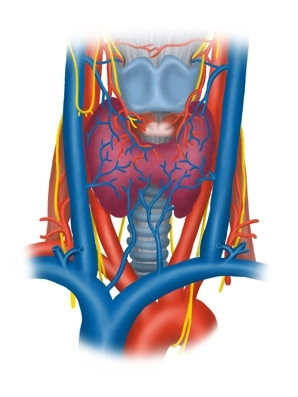
    Before dividing into the internal and external carotid artery, its two main branches, at the superior margin of the thyroid cartilage at the level of its carotid sinus (pressoreceptors for the blood pressure and chemoreceptors for the blood gases), the carotid artery courses in the carotid sheath immediately lateral to the trachea and esophagus. Here, it touches the left and right thyroid lobe as a major blood vessel. The internal jugular vein arises from the sigmoid sinus in the skull, collects the blood from the head and neck, and while coursing caudad it first accompanies the internal carotid artery in the carotid sheath before pursuing a more lateral course, enclosing the lateral aspects of the common carotid artery and vagus nerve (CN X).

    Nerves

    The ansa cervicalis (superior and inferior roots, from C1-C3), which innervates these three above muscles of the anterior triangle of the neck, and the transverse nerve of the neck (from C2/3, innervation of skin and platysma) courses cephalocaudad lateral to the thyroid and next to the vagus nerve and its superior branch to the larynx (superior laryngeal nerve → anterior cricothyroid muscle and mucosa of the superior laryngeal half).

    Fascial layers

    The skin of the anterior triangle of the neck covers several fascial layers (all belonging to the cervical fascia) with distinctive features:

    • The superficial lamina invests all structures of the neck, except for the platysma, and separately invests the sternocleidomastoid muscle as well as the posterior aspect of the trapezius muscle (accessory nerve XI),
    • with the medial pretracheal lamina investing the infrahyoid muscles and
    • the deep prevertebral lamina coursing outside the surgical field between the esophagus and spine.

    Just like the lateral vascular and nerve pedicle (carotid artery, internal jugular vein and vagus nerve), the trachea and thyroid / parathyroids also have their own organ fascias. With their three-dimensional configuration, the fascias invest compartments interspersed with spaces which extend into the mediastinum and thus represent potential routes of infection.

  2. Thyroid region

    Thyroid region

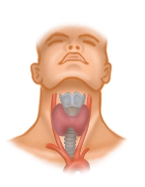
    The unpaired thyroid (glandula thyroidea) and the 4 parathyroids (glandula parathyroidea) are part of the endocrine organ system. The thyroid gland is located in the anterior neck lateral and inferior to the thyroid cartilage, and comprises a left and right lobe and the isthmus which bridges both lobes. The lobes cover the lateral superior rings of the trachea as well as the anterior aspect of both the cricoid and thyroid cartilage; the isthmus sits in the midline at the level of the second and third tracheal ring. The organ is invested by its own capsule of connective tissue (capsula fibrosa) and covered anterolaterally by the infrahyoid muscles (sternohyoid, sternothyroid and omohyoid) and the pretracheal lamina. Due to its embryology and descent (via the thyroglossal duct) from the base of the tongue (foramen caecum) to its later location, its path may leave cysts or ducts to the foramen caecum, as well functional glandular tissue (e.g., pyramidal lobe).

    Blood supply

    Arteries from the external carotid (→ superior thyroid artery) and the subclavian artery via the thyrocervical trunk (→ inferior thyroid artery) with their small anterior and lateral/posterior branches supply the thyroid gland with blood. The thyroidea ima artery, an inconsistent but noteworthy artery arising directly from the aortic arch or the brachiocephalic trunk terminates in the thyroid from below. Venous drainage from this endocrine organ is via corresponding veins (superior and medial thyroid vein → internal jugular vein, inferior thyroid vein → brachiocephalic vein) and has systemic significance.

    Lymphatics and nerves

    Lymphatic drainage from the thyroid is paratracheal and to the deep cervical lymph nodes. Particular attention must be paid to the recurrent laryngeal nerve (from the vagus nerve (CN X), on right: looping around the subclavian artery; on left: looping around the aortic arch). It travels cephalad in the groove between trachea and esophagus to the larynx, where it supplies the sensory mucosal innervation of the inferior half of the larynx and all laryngeal muscles except the anterior cricothyroid (“anticus”).

    The four parathyroids (glandula parathyroidea), small oval glands on the posterior aspect of the thyroid lobes, are supplied by the inferior thyroid arteries.

Topography

Due to their proximity, thyroid surgery runs the risk of injuring not only structures such as the r

Activate now and continue learning straight away.

Single Access

Activation of this course for 3 days.

€7.99 inclusive VAT

Most popular offer

webop - Savings Flex

Combine our learning modules flexibly and save up to 50%.

from €3.29 / module

€39.50 / yearly payment

price overview

general and visceral surgery

Unlock all courses in this module.

€12.42 / month

€149.00 / yearly payment