
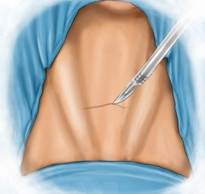
Typical skin incision two fingerbreadths above the suprasternal notch between the bellies of the sternocleidomastoid muscle. Division of subcutaneous tissue and platysma up to the anterior cervical fascia with a sealing instrument. Exposure of the avascular layer between platysma and anterior cervical fascia and mobilization of the skin-platysma flap cranially and caudally.
-
Kocher's collar incision
![Kocher's collar incision]()
Soundsettings -
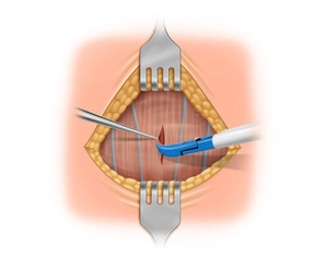
Longitudinal splitting of the straight neck muscles
![Longitudinal splitting of the straight neck muscles]()
Soundsettings Longitudinal splitting of fascia and straight neck muscles in the midline while sparing the superficial neck veins. Then retracting the straight neck muscles from the anterior thyroid capsule.
-
Mobilization of the upper right thyroid pole
![Mobilization of the upper right thyroid pole]()
Soundsettings Approach to the right thyroid side. Right-sided preparation with successive development of the upper pole. Identification and thyroid-adjacent transection of the upper pole vessels after placement of a central clip with the sealing device.
-
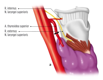
Locating the vagus nerve
![Locating the vagus nerve]()
Soundsettings Careful medialization of the right thyroid lobe with division of the middle thyroid vein (Kocher vein). Locating and looping of the vagus nerve. A normal signal is shown with the neuromonitoring system.
-
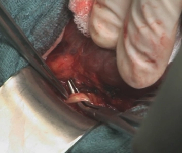
Removal of the right upper parathyroid gland
![Removal of the right upper parathyroid gland]()
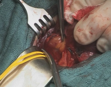
Soundsettings Locating the recurrent laryngeal nerve and identifying it with neuromonitoring. Subsequently exposing the right upper parathyroid gland at its typical location dorsal to the recurrent laryngeal nerve. Excision of the parathyroid gland after placing clips and cauterizing smaller vessels.
Exploration of the inferior parathyroid gland in the normal localization area
After removal of the nodularly enlarged right thyroid lobe (not shown), re-visualization and neurom
After removal of the nodularly enlarged right thyroid lobe (not shown), re-visualization and neurom
Activate now and continue learning straight away.
Single Access
Activation of this course for 3 days.
US$9.30
inclusive VAT
Most popular offer
webop - Savings Flex
Combine our learning modules flexibly and save up to 50%.
from US$7.23 / module
US$86.85/ yearly payment
general and visceral surgery
Unlock all courses in this module.
US$14.47
/ month
US$173.70 / yearly payment
Webop is committed to education. That's why we offer all our content at a fair student rate.