Start your free 3-day trial — no credit card required, full access included

Perioperative management - Aortobifemoral bypass for peripheral arterial disease Fontaine stage IIb–III

  1. Indications

    Depending on the length of the local occlusion/stenosis, the TASC III criteria suggest the treatment options to be used. The length of the stenosis and the region where it is located determine the general type of management: Endovascular or open vascular surgery.

    TASC II classification in aortoiliac occlusion

    Lesion typeMorphologyRecommended treatment

    A

    Short (<3 cm) unilateral or bilateral stenosis of the CIA or EIA  Endovascular

    B

    Single or multiple stenosis totaling 3–10 cm involving the EIA, not extending into the CFA; and/or unilateral CIA occlusionEndovascular

    C

    Bilateral CIA occlusion;[MM3]  bilateral EIA stenoses 3–10 cm long not extending into the CFA; unilateral complete EIA occlusion Open reconstruction

    D

    Diffuse disease involving the aorta and both iliac arteries requiring treatment; unilateral occlusions of both CIA and EIA; bilateral occlusions of EIA  Open reconstruction


    Video example

    Präoperatives Angiogramm
    Preoperative angiogram


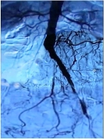
    Clinically the patient is classified as Fontaine PAOD IIb–III and the preoperative angiogram demonstrates: Occlusion of the right CIA and EIA, subtotal stenosis of the left EIA and bilateral stenosis of the femoral bifurcation

    -> TASC D, , thus recommendation for  open revascularization

    PAOD classification according to Fontaine and Rutherford


    Fontaine stage

    Clinical symptoms                                    

    Rutherford

    Grade

    Clinical symptoms

    I

    Asymptomatic

    0

    0

    Asymptomatic

    IIa

    Distance > 200 m

    1

    I

    Mild claudication

    IIb

    Distance< 200 m

    2

    I

    Moderate claudication


    3

    I

    Severe claudication

    III

    Ischemic rest pain

    4

    II

    Ischemic rest pain

    IV

    Ulcers or gangrene

    5

    III

    Ischemic ulceration not exceeding ulcer of the digits of the foot
      

    6

    III

    Severe ischemic ulcers or frank gangrene
  2. Contraindications

    • Serious cardiopulmonary risks (e.g., NYHA IV, COPD GOLD stage IV)
    • Acute or chronic inflammatory abdominal disease (e.g., florid ulcerative colitis, recurrent sigmoid diverticulitis)
    • History of multiple extensive abdominal procedures ("hostile abdomen")
    • Cirrhosis
    • Advanced malignancy
  3. Preoperative diagnostic work-up

    Medical history

    • Claudication
    • Distance walking without complaints
    • Risk factors -> nicotine; arterial hypertension; CHD; heart failure; diabetes; manifest renal failure with/without dialysis; coagulation disorder

    Inspection

    • Skin changes
    • Muscular abnormalities
    • Orthopedic deformities
    • Skin color
    • Hair
    • Trophic changes
    • Swelling; edema; mycosis; phlegmon; leg ulcers etc.    

    Palpation with contralateral comparison

    • Arterial pulse examination
    • Skin temperature

    Arterial auscultation of the extremities with contralateral comparison

    Palpation-Auskultation
    Palpation–auscultation


    Ankle-Brachial Index (ABI)

    • ABI = systolic BP of posterior tibial artery / systolic BP of brachial artery
    ABISeverity of PAOD
    > 1.3false high values (suspected Mönckeberg arteriosclerosis, e.g. in diabetes)
    > 0.9Normal finding
    0.75 - 0.9Mild PAOD
    0.5 - 0.75Moderate PAOD
    < 0.5Severe PAOD
    • An ABI of < 0.9 demonstrates the presence of significant PAOD.
    • Noninvasive Doppler ultrasound measurement of the Ankle-Brachial Index (ABI) is an adequate test for the presence of PAOD.
    • For the diagnosis of PAOD, the ABI with the lowest posterior tibial artery pressure is used.
    • A pathologic ankle-arm index is an independent risk indicator for increased cardiovascular morbidity and mortality.

    Color coded duplex sonography

    • Carotid arteries, abdominal aorta, arteries of the extremities
    • Localization of stenoses and occlusions in almost all vascular regions except the chest
    • Quantifies the degree of stenosis and allows assessment of plaque morphology
    • Sensitivity and specificity about 90%.   
    • Well suited for screening purposes

    CT angiography

    • Multi-slice computed tomography (MS-CT) with non-ionic contrast medium
    • Broad range of indications: Traumatic vascular lesion (especially trunk); vascular dissection/rupture; aneurysm; arterial thrombosis/embolism; portal vein/mesenteric vein thrombosis; pulmonary artery embolism; PAOD; vascular tumors
    • Pros: Fast; detection of significant concomitant disease; imaging of peripheral arteries; sensitivity and specificity each about 90%.
    • Cons: Exposure to radiation and contrast media, allergies ( about 3%), no functional assessment

    Cardiac check-up

    • Resting ECG
    • Exercise ECG
    • Echocardiography

    Chest radiograph

    Possibly spirometry

    Clinical chemistry

    • RBC
    • Electrolytes
    • Coagulation
    • Renal function
    • Liver function
    • Blood lipids
    • Blood group
  4. Special preparation

    • Enema evening before surgery
    • Hair trimmed in surgical field
    • Packed RBCs ordered
    • Foley catheter
    • Perioperative prophylactic antibiotics 30 minutes before surgery (see German KRINKO and Robert-Koch-Institut recommendations)
  5. Informed consent

    General surgical risks

    • Major bleeding; blood transfusions; hepatitis/HIV transmission from allogeneic blood units.
    • Allergy/incompatibility
    • Wound infection
    • Thrombosis/embolism
    • Injury to skin, vessels, nerves, e.g. due to positioning.
    • Keloids
    • Incisional hernia

    Specific surgical risks

    • Thrombosis of grafts and possibly regions supplied by runoff vessels, possibly leg ischemia, amputation
    • Graft infection with suture bleeding; sepsis; leg ischemia; amputation
    • Injury to adjacent organs such as ureter, bladder, spleen, liver; intestinal ischemia -> resection, colo-/ileostomy
    • Paraplegia in artery of Adamkiewicz with low origin
    • Nerve lesions -> paresthesia; pain; paralysis of the abdominal wall and thigh muscles
    • Peritoneal adhesions -> chronic pain, mechanical ileus
    • Lymphatic fistula
    • Secondary bleeding
    • Impotence    
    • Anastomotic/graft aneurysm
    • Impaired renal function by intraoperative angiography
  6. Anesthesia

  7. Positioning

    Positioning

    Supine position, left arm abducted, right arm adducted. The upper body should be slightly reclined at the level of the thoracoabdominal transition between pubic bone and xyphoid to increase the distance between iliac crest and costal margin

  8. Operating room setup

    Operating room setup

    The surgeon is on the right side of the patient facing the first assistant on the left side. The second assistant is to the surgeon’s left. The scrub nurse is to the left of the first assistant.

  9. Special instruments and fixation systems

    • Abdominal wall retractor system
    • Aortic cross-clamps, bulldog clamps
    • Inverted Y-grafts of different sizes (14–22 mm)
    • Nonabsorbable monofilament vascular sutures (3-0 to 6-0)
    • Cell salvage
  10. Postoperative management

    Postoperative analgesia

    Follow these links to Prospect (Procedures Specific Postoperative Pain Management) and the current German guideline Leitlinien der Behandlung akuter perioperativer und posttraumatischer Schmerzen [Guidelines on treatment of acute perioperative and posttraumatic pain].

    Postoperative management:

    • 24-hour recovery on ICU, possibly on IMCU
    • If possible regular ward from postop. day 1–3
    • Close monitoring of CV system and lungs
    • Monitoring of leg pulses, capillary perfusion of the feet

    Deep venous thrombosis prophylaxis:

    • Low molecular weight heparin by weight; in multifocal arteriosclerosis ASA 100 mg/d q.d. on return to nutrition

    Ambulation

    • Edge of bed on day 2 (caution: inguinal anastomosis: rupture on forced mobilization possible)

    Physical therapy

    • Isometric and breathing exercises    

    Diet

    • Gradual return to regular nutrition after initial bowel movement      

    Bowel movement:

    • Enema in case of no spontaneous bowel movement by day 3
    • Gastrografin if unsuccessful
    • Neostigmin as a means of last resort

    Work disability

    • About 3 months