VATS Thymectomy

Reading time readingtime 08:28 min.
Activate now
  1. Placement of accesses for VATS thymectomy

    Video
    Placement of accesses for VATS thymectomy

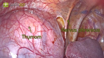
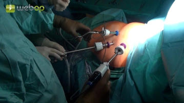
    The video-assisted thoracoscopic thymectomy is presented here using a 3-port technique with CO2 insufflation. During operations on the mediastinum, CO2 insufflation significantly facilitates layer-specific dissection, so trocars with appropriate valves and gas connections, as used in visceral surgery. The first access is created via a mini-incision in the area of approximately the 5th intercostal space, anterior axillary line, in extension of the inframammary fold. The placement of the 2 additional incisions should only be performed after inspection of the site. There are various possibilities here. Usually, a trocar in the 3rd intercostal space in the anterior axillary line and another a few centimeters ventral to the first trocar in the same intercostal space are suitable for the additional accesses. Alternatively, a substernal trocar is also possible, which can be more easily expanded for later retrieval in the case of larger tumors.

    Note:

    • The thoracoscopic thymectomy can be performed from the left as shown here. Alternatively, access from the right or subxiphoidally is also possible.
    • Especially in obese patients, CO2 insufflation can be advantageous.
  2. Visualization of the phrenic nerve and determination of the lateral resection margin

    Video
    Visualization of the phrenic nerve and determination of the lateral resection margin

    First, the pleural covering at the mediastinum is gradually opened with the endoscopic dissecting scissors. Subsequently, the phrenic nerve must be clearly identified. The nerve forms the lateral resection margin. Through CO2 insufflation, the tissue planes become clearly visible after incision of the pleura, allowing for bloodless and layer-appropriate dissection.

  3. Circular preparation of the mediastinal pleura

    Video
    Circular preparation of the mediastinal pleura

    After clearly visualizing the phrenic nerve, further dissection can be continued with a high-energy device to prevent smaller bleedings, which lead to poorer visibility, or later lymphatic fistulas. The dissection is now continued circumferentially around the thymus.

  4. Visualization of the left thymic horn

    Video
    Visualization of the left thymic horn

    Now follows the visualization of the left thymic horn. Here are also located the thymic veins, which drain blood from the thymus into the left brachiocephalic vein (=vena anonyma). The thymic veins are occasionally so weakly developed that they can be safely transected using a high energy device. Otherwise, clipping is advisable. During dissection, always pay attention to the brachiocephalic vein.

Resection of the thymoma from the pericardium

After preparation and exposure of the cranial portion of the tumor, the thymus is now detached from

Activate now and continue learning straight away.

Single Access

Activation of this course for 3 days.

€7.99 inclusive VAT

Most popular offer

webop - Savings Flex

Combine our learning modules flexibly and save up to 50%.

from €3.29 / module

€39.50 / yearly payment

price overview

thoracic

Unlock all courses in this module.

€7.42 / month

€89.00 / yearly payment