Anatomy - Fundoplication, Short-Floppy-Nissen technique, hiatal repair with mesh augmentation

  1. Surgical Anatomy of the Stomach

    Surgical Anatomy of the Stomach

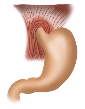
    The stomach is, formally speaking, a dilation of the digestive tract located between the esophagus and the intestine, tasked with storing and mixing food. This muscular hollow organ produces acidic gastric juice (mucus and HCl) and enzymes that partially digest some components of food, subsequently transferring the chyme in portions to the small intestine.

    The stomach is usually located in the left and middle upper abdomen directly beneath the diaphragm. The position, size, and shape of the stomach vary greatly from person to person and depending on age, state of fullness, and body position. When moderately filled, the stomach is on average 25-30 cm long and has a storage capacity of 1.5 liters, and in extreme cases, up to 2.5 liters.

    The stomach is anchored and stabilized in the abdominal cavity by ligaments that extend to the liver and spleen, among others. It forms the greater curvature (Curvatura major) with its convex side and the lesser curvature (Curvatura minor) with its concave side. Its anterior wall is referred to as Paries anterior, and its posterior wall as Paries posterior.

    The stomach is intraperitoneal and thus covered with serosa, except for the dorsal cardia, which is free of serosa. The embryonic mesogastria move from their former sagittal position to a frontal one through gastric rotation: The lesser omentum extends from the lesser curvature to the liver hilum, while the greater omentum spreads from the greater curvature to the transverse colon, spleen, and diaphragm.

    The stomach can be divided into different sections:

    Cardia / Ostium cardiacum
    The upper stomach mouth is an area of 1-2 cm where the esophagus opens into the stomach. Here is the sharp transition from the esophageal mucosa to the gastric mucosa, which is usually easily recognizable with an endoscope.

    Fundus gastricus
    Above the cardia, the fundus arches upward, also known as the "gastric dome" or Fornix gastricus. The fundus is typically filled with air that is involuntarily swallowed while eating. In an upright person, the fundus forms the highest point of the stomach, so in an X-ray, the collected air appears as a "gastric bubble." Opposite the cardia, the fundus is demarcated by a sharp fold (Incisura cardialis).

    Corpus gastricum
    The main part of the stomach is formed by the corpus. Here, deep longitudinal folds of the mucosa (Plicae gastricae) extend from the cardia to the pylorus and are also referred to as the "gastric street."

    Pars pylorica
    This section begins with the expanded antrum pyloricum, followed by the pyloric canal (Canalis pyloricus), and ends with the actual pylorus. Here lies the pyloric sphincter (M. sphincter pylori), formed by a strong circular muscle layer, which closes the lower stomach mouth (Ostium pyloricum). The pylorus closes the stomach exit and periodically allows some chyme to pass into the subsequent duodenum.
  2. Layers and Structure of the Stomach Wall

    Stomach Wall

    Under the microscope, the stomach wall shows a characteristic layer structure from inside to outside:

    • Inside, the stomach wall is lined by mucosa (Tunica mucosa). The gastric mucosa is divided into three sublayers: The lamina epithelialis mucosae produces a viscous neutral mucus that protects the gastric mucosa from mechanical, thermal, and enzymatic damage. Below it is the lamina propria mucosae, which serves as a shifting layer and contains the gastric glands (Glandulae gastricae). Finally, there is a narrow lamina muscularis mucosae that can alter the relief of the mucosa.
    • The gastric mucosa is followed by a loose shifting layer (Tela submucosa) consisting of connective tissue, through which runs a dense network of blood and lymph vessels, as well as a nerve fiber network, the submucosal plexus (Meissner's plexus), which controls gastric secretion. This plexus operates independently of the central nervous system (CNS) but can be influenced by it via the autonomic nervous system.
    • Next is a strong tunica muscularis, divided into three sublayers with fibers running in different directions: an inner layer of small obliquely running muscle fibers (Fibrae obliquae), then a circular muscle layer (Stratum circulare), and an outer longitudinal muscle layer (Stratum longitudinale). This musculature is responsible for the peristalsis of the stomach, which is essential for the continuous mixing of the chyme with gastric juice. Between the circular and longitudinal muscle layers runs a nerve fiber network, the myenteric plexus (Auerbach's plexus), which controls the function of the musculature. Like the submucosal plexus, this plexus operates largely autonomously but is influenced by the autonomic nervous system.
    • Another connective tissue shifting layer (Tela subserosa) follows.
    • The peritoneum as the abdominal lining (Tunica serosa) forms the conclusion.

    Gastric Glands

    The gastric glands (Glandulae gastricae) are located in the lamina propria mucosae and can be found in the fundus and corpus of the stomach. Up to 100 glands are located on 1 mm² of the mucosal surface. Various cells are located in the wall of the glandular tube:

    • Mucous cells: They produce the same neutral mucus as the epithelial cells.
    • Neck cells: These cells are located quite superficially in the gland and secrete alkaline mucus, i.e., the pH value is high due to the bicarbonate ions (OH ions) contained in it. This property is important to control and, if necessary, regulate the pH value of the stomach. The mucus coats the gastric mucosa and thus protects against self-digestion by the aggressive hydrochloric acid (HCl) and enzymes as self-digesting proteins. This cell type is found predominantly in the cardia and fundus of the stomach.
    • Chief cells: These cells produce the inactive precursor enzyme pepsinogen, which is converted into the active enzyme pepsin by hydrochloric acid (HCl) after release and is responsible for the digestion of dietary proteins. Since the enzyme only comes into contact with hydrochloric acid at the surface of the gland, self-digestion of the glands by the enzyme is prevented. This cell form is mainly located in the corpus of the stomach.
    • Parietal cells: These cells, which are found more frequently in the gastric corpus, produce abundant hydrogen ions (H+ ions) needed for the formation of hydrochloric acid (HCl). Hydrochloric acid has a very low pH value of 0.9-1.5. In addition, parietal cells produce the so-called intrinsic factor. This substance forms a complex with vitamin B12 from food in the intestine, which can then pass through the small intestine wall. This vitamin is of particular importance in erythropoiesis (stomach removal can lead to anemia).
    • G cells: These cells, which are preferably located in the antrum of the stomach, produce gastrin to increase HCl production in the parietal cells.
  3. Function

    The stomach serves as a reservoir for ingested food. It can store food for hours, allowing us to meet our daily nutritional needs with a few larger meals. Through peristalsis, the chyme is mixed with gastric juice, the food is chemically broken down, partially digested, and then gradually transferred to the duodenum.

Arterial, venous, and neural supply

The arterial supply of the stomach is provided by several blood vessels, all originating from the u

Activate now and continue learning straight away.

Single Access

Activation of this course for 3 days.

€7.99 inclusive VAT

Most popular offer

webop - Savings Flex

Combine our learning modules flexibly and save up to 50%.

from €3.70 / module

€44.50 / yearly payment

price overview

general and visceral surgery

Unlock all courses in this module.

€12.42 / month

€149.00 / yearly payment