Perioperative management - Fundoplication, laparoscopic according to Toupet

  1. Indications

    Indications

    A hiatal hernia occurs due to a weakening of the diaphragm, where varying portions of the stomach, up to the entire stomach in severe cases, along with the spleen and intestinal parts, may shift into the thoracic cavity through a gap between the diaphragm's crura.

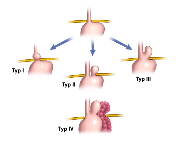
    There are 4 types distinguished

    Type I: Axial hernia, meaning the stomach entrance moves along the organ's axis through the hiatus upwards.

    Type II: Paraesophageal hernia, meaning the stomach entrance remains below the diaphragm while other parts of the stomach move alongside the esophagus through the hiatus into the thoracic cavity.

    Type III: Mixed form of Type I and II. There is both an axial shift of the esophagus and stomach towards the thoracic cavity and a displacement of stomach parts up to the entire stomach alongside the esophagus (thoracic stomach – "upside-down stomach").

    Type IV: The most severe form, where additionally parts of the colon, the spleen with or without the pancreatic tail, are displaced into the thoracic cavity.

    Axial sliding hernias account for up to 90% of cases and are initially described as a normal variant rather than a disease per se.

    The indication for surgery depends on the symptomatology and hernia morphology.

    While the goal of antireflux surgery is to strengthen the lower esophageal sphincter with a gastric wrap to prevent reflux, the goal for hiatal hernias of Types II-IV is to correct the incorrect position of the stomach and other organs displaced into the thoracic cavity.

    An indication for surgery exists for:

    • Axial hiatal hernia with therapy-resistant reflux disease

    Note: An antireflux operation should only be performed if there is a long-term need for therapy (>1 year).

    • Complicated GERD ("gastroesophageal reflux disease"): severe reflux esophagitis (Los Angeles Grade C and D), peptic stricture

    The Los Angeles classification is the most commonly used grading system for reflux esophagitis. The Los Angeles classification is based on the endoscopic examination of the esophagus. It distinguishes 4 stages:

    • Stage A: One or more mucosal lesions < 0.5 cm. The lesions do not extend beyond the tops of two mucosal folds.
    • Stage B: At least one lesion > 0.5 cm. However, the lesions do not yet extend beyond the tops of two mucosal folds.
    • Stage C: The lesions extend beyond the tops of several (>2) mucosal folds. However, they occupy less than 75% of the total circumference of the esophagus, i.e., there are no circular defects yet.
    • Stage D: There are circular lesions that occupy more than 75% of the total circumference of the esophagus.


    • Disease not completely treatable with medication

    Note: An initial response to PPIs with increasing resistance indicates the success of the operation.

    • Patient's desire not to take proton pump inhibitors for life.
    • Accompanying hiatal hernia Type II to IV

    Note: For hiatal hernias Type II to IV, the hernia itself is already an indication for surgery due to the possibility of severe complications (incarceration).

    The operation of the hiatal hernia includes repositioning the herniated content and a hiatoplasty (narrowing of the esophageal hiatus) by suture. Depending on the clinical situation, the procedure is supplemented by sac removal, fundoplication, mesh implantation, or gastropexy.

    The laparoscopic or robotically assisted transabdominal approach is nowadays the preferred method for treating hiatal hernia.

    The surgical treatment of reflux disease requires a fundoplication in addition to the treatment of the hiatus.

    Minimally invasive vs. open

    The superiority of laparoscopic fundoplication over open fundoplication is clearly documented in the literature. The guideline explicitly demands that the laparoscopic technique should be used as the standard.

    Type of wrap

    It has long been controversially discussed whether the partial posterior fundoplication according to Toupet is superior to the total fundoplication according to Nissen in the treatment of GERD. Various studies have attempted to resolve the "Toupet vs. Nissen" controversy.

    The full wrap seems to be associated with a higher incidence of postoperative dysphagia and "gas bloating," while persistent reflux symptoms are more common with a partial wrap. In summary, the available data tends to favor the superiority of the 270° Toupet wrap for the treatment of gastroesophageal reflux disease.

    Dysphagia

    The dysphagia issue has been improved in recent years through the possibilities of endoscopic bougienage. Good results are already seen after 1–2 sessions with large-caliber bougies. In fact, the frequency of reoperations due to persistent dysphagia is rather low.

    Sac removal

    It is recommended to dissect the hernia sac from the mediastinal structures, which is (not clearly proven) associated with a lower recurrence rate, although it is not always possible with large hernias due to the high risk of iatrogenic injury to the esophagus or gastric vessels.

  2. Contraindications

    Special:

    • Esophageal motility disorders (achalasia, diffuse esophageal spasm)
    • Esophageal involvement in an autoimmune disease, e.g., CREST syndrome as a special form of scleroderma (C – Calcinosis, R – Raynaud's phenomenon, E - Esophageal dysfunction, S – Sclerodactyly, T – Telangiectasia)
    • Esophagitis of other origins (infectious, drug-induced toxic)
    • Extraesophageal diseases with reflux-like symptoms, e.g., coronary heart disease

    Note: Special caution is advised for patients whose symptoms of a presumed reflux disease do not or only insignificantly respond to high-dose therapy with PPIs. In such cases, the diagnosis of reflux disease should be critically reviewed, and it should be clarified why the drug treatment was not successful. The failure of conservative treatment does not constitute an immediate indication for surgery; a misdiagnosis should always be considered.

    General:

    • Contraindications for the creation of a pneumoperitoneum due to severe systemic disease or massive adhesions ("hostile abdomen").

    Additionally, relative contraindications should be considered, where preoperative optimization may be possible:

    • Severe coagulation disorders (Quick < 50%, PTT > 60 sec, platelets < 50/nl)
    • Severe portal hypertension with caput medusae
    • Patients with severe cardiovascular comorbidities for whom anesthesia alone poses a risk (e.g., NYHA III/IV constellation, high-grade carotid stenoses).
  3. Preoperative Diagnostics

    Medical History:

    • Reflux symptoms: heartburn, acid regurgitation, regurgitation, feeling of pressure
    • Long-standing history of reflux
    • Positive PPI response
    • Necessary increase in PPI dosage/ PPI intolerance/ unwillingness to take PPI
    • Reduced quality of life / intolerable reflux symptoms
    • The patient's distress is to be assessed with a Quality of Life Index.
    • Recurrent aspirations

    PPI Test:

    The complete or significant response to a PPI administration indicates the presence of a reflux disease, or in other words: if a high-dose PPI therapy fails, the presence of GERD is rather unlikely.

    The PPI test is only meaningful in the presence of symptoms that already suggest reflux disease, while endoscopy is unremarkable. The PPI test should be conducted with two to three times the standard dosage recommended for reflux treatment over at least 2 weeks, as reflux episodes can vary from day to day or occur intermittently without therapy.

    Gastroscopy: 

    The importance of an esophagogastroduodenoscopy in reflux diagnostics is undisputed and mandatory before surgical intervention.

    It enables:

    • Diagnosis of reflux esophagitis and assessment of its severity (also as follow-up control in esophagitis therapy)
    • Diagnosis of a hiatal hernia
    • Detection of complications (stricture, ulcer)
    • Exclusion of malignancy

    Early endoscopy is indicated in unusually severe symptoms and alarm symptoms such as anemia, dysphagia, and weight loss. Obtaining a histology is mandatory for all macroscopic abnormalities.

    The so-called "Z-line" (transition of the squamous epithelium of the esophagus into the columnar epithelium of the stomach) serves as a guide structure, which normally lies exactly at the hiatus. In an axial hernia, this line moves cranially. Depending on the distance from the diaphragm passage, it is referred to as a small (< 3cm) or large hernia(> 3cm).

    Large hiatal hernias can lead to intraluminal bleeding due to trauma to the mobile stomach. Linear ulcerations of the stomach at the level of the hiatus (Cameron lesions) with chronic anemia can occur.

    In a paraesophageal hernia, the hernia appears beside the non-displaced esophagus during endoscopic inversion. Asymptomatic hiatal hernias are usually incidental findings.

    A reflux esophagitis is always endoscopically classified before invasive therapy. The Los Angeles Classification should be used for this, see point 1 (Indications).

    24-Hour Esophageal pH Monitoring: 

    The 24-hour esophageal pH monitoring is the gold standard for objectifying gastroesophageal reflux.

    It records the circadian rhythm of reflux episodes, physical activities, food intake, and body positions. A symptom correlation with the recorded reflux episodes through patient documentation increases the sensitivity of the pH monitoring.

    DeMeester Score: Evaluation system for quantifying gastroesophageal reflux episodes through a 24-hour long-term pH monitoring. The score considers the pH value in the distal esophagus as well as the frequency and duration of reflux. The most important parameter is the percentage of time at a pH value < 4. A DeMeester Score ≥ 14.72 is pathological.

    A 24-hour pH monitoring is indicated for:

    • Preoperative documentation of pathological reflux
    • Persistence of reflux symptoms under adequate PPI medication
    • In endoscopically unremarkable "NERD" patients (= Non Erosive Reflux Disease)
    • Recurrent reflux symptoms after antireflux surgery

    In pH monitoring, it must be noted that up to 25% of patients with reflux esophagitis and around 30% of NERD patients have normal values, which is due to the fact that even with clear reflux disease, the amount of reflux can vary from day to day.

    Esophageal Manometry:

    Examination to assess pressure and functional processes of the esophagus at rest and during swallowing. It is used for diagnosing esophageal motility disorders (both hypomotile and hypermotile).

    Esophageal manometry can reliably assess the competence of the lower sphincter (resting pressure, length) and the tubular motility of the esophagus. The value of manometry for the primary diagnosis of GERD is low, but it can be useful in individual cases to differentiate other motility disorders of the esophagus (e.g., achalasia).

    It is highly recommended in the context of preoperative evaluation and documentation regarding the selection of the surgical procedure. In the presence of tubular contraction disorders or achalasia, a Toupet or Nissen fundoplication is contraindicated. 

    Radiological Procedures:

    Radiological procedures can support the diagnosis of a hiatal hernia or other therapy-relevant issues.

    In a chest X-ray, a mirror image in the herniated stomach can be detected.

    An esophageal barium swallow examination can contribute to further diagnostic clarification, especially for detecting a hiatal hernia and differentiating the type of hernia, but it has no significance for diagnosing reflux disease. Reflux is physiological, so no disease value can be derived from the radiological representation. Furthermore, reflux occurs intermittently and can only be reliably determined through long-term measurement, not through a radiological snapshot.

    Many surgeons still find the barium swallow helpful to visualize the anatomy of the gastroesophageal junction before a planned operation. Also, in the clarification of postoperative problems after a fundoplication, the barium swallow is often indispensable, as, for example, the resolution of the wrap and a telescoping phenomenon can hardly be assessed endoscopically.

    For comprehensive imaging, an MRI or CT is always additionally used, especially in type II-IV hernias.

    The Montreal Classification of Gastroesophageal Reflux Disease (GERD)

    Since 2006, the different manifestations of GERD have been clearly represented by the so-called Montreal Classification.

    Initially, the Montreal Classification provides a well-suited definition of GERD as the backflow (reflux) of stomach contents into the esophagus, which leads to symptoms and/or complications that manifest esophageally or extraesophageally. The reflux disease is very heterogeneous, meaning the disease can manifest in very different ways. The diagram clearly visualizes the wide range of problems that can arise in the esophagus (esophageal) and other adjacent organs (extraesophageal) due to reflux.

    505-Montreal_Klassifikation


    In type II to IV hernias, clinically relevant reflux is not the leading symptom but rather entrapment phenomena of the herniated organ and passage disorders up to complete gastric obstruction. Even more dangerous is a compromise of blood circulation in the herniated sections with the possible development of necrosis and consequent hollow organ perforation.

  4. Special Preparation

    • Shaving: Nipples to thighs
    • Preoperative nutrition: Regular diet
    • Epidural catheter: not indicated
    • Single-shot antibiotic with Cefuroxime 1.5g intravenously approximately 30 minutes before skin incision.
    • Compression stockings
    • Breathing exercises for COPD or accompanying large hiatal hernia
    • Preoperative review and adjustment of anticoagulant therapy:
      • Perioperative therapy with aspirin can be continued. 
      • Clopidogrel (ADP inhibitor) should be paused at least 5 days prior. 
      • Vitamin K antagonists should be paused 7-10 days under INR control and "bridged" with low molecular weight heparin subcutaneously.
      • NOAC (new oral anticoagulants) should be paused 2-3 days preoperatively
      • In cases of very high closure/insult risk, interdisciplinary therapeutic concept regarding indication of anticoagulation, possibility of bridging with heparin, and operative bleeding risk.
  5. Informed consent

    General:

    • Bleeding/postoperative bleeding with administration of donor blood
    • Thromboembolism
    • Drain insertion, catheter insertion
    • Conversion in case of complications
    • Possible need for surgical/interventional revision due to a complication
    • Wound infection/abscess
    • Trocar hernia

    Specific:

    • Esophageal/stomach injuries
    • Injury to adjacent structures (spleen, pancreas, small intestine, colon, liver, gallbladder)
    • Need for surgical extension
    • Pneumothorax
    • Postoperative dysphagia
    • Gas-bloat syndrome (inability to belch or vomit; note: may also exist preoperatively! History!)
    • Increased retention of air in the gastrointestinal tract
    • Denervation syndrome due to damage to the anterior or posterior vagus branch (consequence: gastric emptying disorder, diarrhea)
    • Lack of success (despite correct indication and surgical technique)
    • Recurrence
Anesthesia

Intubation anesthesia in pneumoperitoneum ... - Operations in general, visceral and transplant surg

Activate now and continue learning straight away.

Single Access

Activation of this course for 3 days.

€7.99 inclusive VAT

Most popular offer

webop - Savings Flex

Combine our learning modules flexibly and save up to 50%.

from €3.70 / module

€44.50 / yearly payment

price overview

general and visceral surgery

Unlock all courses in this module.

€12.42 / month

€149.00 / yearly payment