Start your free 3-day trial — no credit card required, full access included

Perioperative management - Femoral hernia repair, TIPP technique

  1. Indication

    Indication

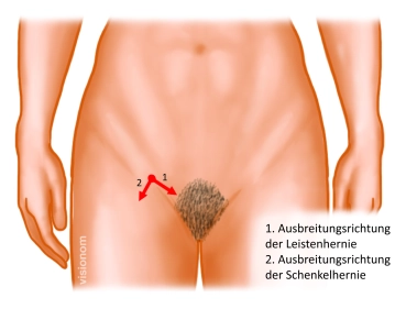
    Femoral hernias incarcerate significantly more often than inguinal hernias. Therefore, the indication for surgical treatment should generally be made. However, no diagnostic procedure can distinguish between inguinal and femoral hernias.

    Femoral hernia occurs significantly more frequently in women than in men, so every inguinal hernia in women should be operated on promptly.

    In the case of femoral hernia, laparoscopic techniques are preferred over open procedures due to less postoperative and chronic pain and the easier placement of the mesh covering all potential hernia orifices. Since ultimately in the described open procedure the mesh is placed in the posterior layer, reoperation is more difficult. The advantage is the lower invasiveness and the possibility to perform the procedure under local anesthesia.

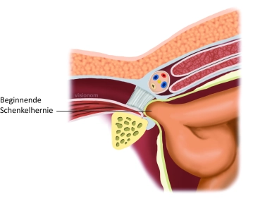
    In incarcerated femoral/inguinal hernias, which are distinguished from irreducible hernias by pronounced pain, acute onset, and signs of bowel obstruction, the diagnostic superiority of laparoscopy should be utilized. Its advantage is the possibility of repositioning the incarceration with subsequent assessment of organ perfusion. In about 90% of cases, organ perfusion recovers after repositioning.

    The treatment of femoral hernia can be performed immediately or at a later time depending on the local infection situation. In principle, a suture closure of the femoral area is possible even if bowel resection is necessary.

  2. Contraindications

    • In cases of severe preoperative pain, preference should be given to minimally invasive procedures.
    • Skin infection in the area of access
    • Due to the possibility of performing the procedure under local anesthesia, there are only a few absolute contraindications:
      • Uncorrectable coagulation disorders
      • Incurable intra-abdominal diseases with a poor prognosis (peritoneal carcinomatosis)
  3. Preoperative Diagnostics

    Preoperative Diagnostics

    Patients with a femoral hernia often experience nonspecific pain in the groin area radiating to the thigh. Swelling is only partially visible or palpable.

    The femoral hernia often presents as an ileus, with a definitive diagnosis of a femoral hernia due to lack of palpability only being made intraoperatively during exploration.

    If the bladder wall is part of the hernia contents or is mechanically irritated by the hernia, dysuria and hematuria may occur.

    In cases of incarceration, redness, swelling, pain, and symptoms of an ileus occur.

    • Differential diagnoses

    other masses: lymphadenitis, lipoma, femoral artery aneurysm, varix node, other tumors

    other causes of pain: groin strain, adductor syndrome, nerve irritation, etc.

    Ultrasound is used to confirm the diagnosis, and in exceptional cases, a computed tomography (CT) scan.

  4. Special Preparation

    • Clear marking of the side on the skin of the awake patient before possibly taking premedication.
    • Perioperative antibiotic prophylaxis is recommended for open procedures with mesh, especially with an increased risk profile.

  5. Informed consent

    • Information about alternative procedures
    • Mesh implantation/mesh infection
    • Handling of nerves, possibly neurectomy
    • Injury to the vas deferens and testicular vessels
    • Persistent sensory disturbance (20%)
    • Severe chronic pain (3%) risk dependent on preoperative symptoms
    • Recurrence (2%)

    General complications:

    • Seroma
    • Hematoma
    • Wound healing disorder
    • Infection
    • Thrombosis and embolism
    • Injuries to, for example, nerves, vessels, bowel, bladder
    • (Post-)bleeding
    • Follow-up surgery
    • Bowel resection
    • Lethality


  6. Anesthesia

  7. Positioning

    Positioning

    Supine position with both arms ideally positioned outwards

  8. OR Setup

    OR Setup
    • The surgeon stands on the side to be operated on
    • The first assistant stands on the opposite side
    • The scrub nurse stands to the left of the assistant and has the instrument table at the foot end on the surgeon's side
  9. Special Instruments and Retention Systems

    • Basic Tray
    • Non-resorbable, large-pore, surface-reduced synthetic mesh
  10. Postoperative Treatment

    Postoperative Analgesia: Systemic analgesics as needed. Follow the link here to PROSPECT (Procedures Specific Postoperative Pain Management).

    Follow the link here to the current guideline: Treatment of acute perioperative and post-traumatic pain.

    Medical Follow-up: Removal of the Redon drain – if present – usually after 24 hours
    Thrombosis Prophylaxis: Thrombosis prophylaxis with low molecular weight heparin in prophylactic, possibly weight-adjusted dosage depending on individual thrombosis risk.
    Follow the link here to the current guideline: Prophylaxis of venous thromboembolism (VTE).

    Note: Renal function, HIT II (history, platelet control)

    Mobilization: immediate mobilization
    Physical Therapy: breathing exercises for pneumonia prophylaxis only in bedridden patients
    Diet Progression: rapid diet progression
    Bowel Regulation: prevention of constipation, if necessary, laxatives from the 2nd day
    Incapacity for Work: incapacity for work 7 to 14 days, depending on occupational physical strain