
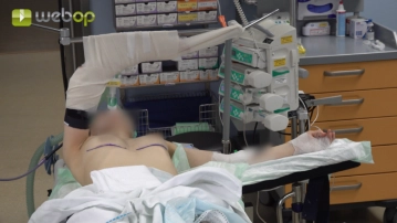
In the supine position, the left arm is extended, and the right arm is fixed at 90° flexion at the elbow joint on the support. The right shoulder and back are padded for stabilization.
-
Positioning
![Positioning]()
-
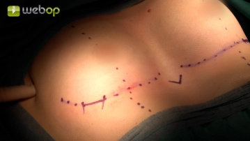
Definition of topographical landmarks
![Definition of topographical landmarks]()
Soundsettings Preoperatively, the inframammary fold was marked while standing. Now, the nipple/medioclavicular lines are added. Subsequently, the xiphoid process is palpated and also marked. This is followed by identifying the planned entry points for the pectus bar laterally, at the level of the highest point of the deformity. These are marked on the inframammary lines.
-
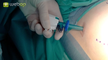
Insertion of the optical trocar
![Insertion of the optical trocar]()
Soundsettings Through a small skin incision on the mid-axillary line at the level of the axilla, the optical trocar is inserted into the right thorax under apnea. After blocking the trocar to prevent dislocation, CO2 insufflation is connected and the camera with 30° optics is introduced. Explorative thoracoscopy.
Preparation of the entry points
First, an incision is made at the previously determined sites using a scalpel. Then, preparation is
First, an incision is made at the previously determined sites using a scalpel. Then, preparation is
Activate now and continue learning straight away.
Single Access
Activation of this course for 3 days.
US$9.10
inclusive VAT
Most popular offer
webop - Savings Flex
Combine our learning modules flexibly and save up to 50%.
from US$4.23 / module
US$50.80/ yearly payment
thoracic
Unlock all courses in this module.
US$8.46
/ month
US$101.60 / yearly payment
Webop is committed to education. That's why we offer all our content at a fair student rate.