
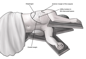
Incision of approximately 4 cm in length in the area of the anterior axillary line at the upper edge of the 5th rib to access the 4th intercostal space above. As a guide, an imaginary line from the tip of the scapula to the nipple is often helpful. Transection of the subcutis on the rib with the monopolar knife. Subsequently, stepwise preparation of the intercostal muscles with the monopolar knife. The pleura is opened bluntly with a finger. Palpation of the thoracic wall for adhesions and insertion of a wound protection film.
-
Access uniportal VATS right
![Access uniportal VATS right]()
-
Visualization of the pulmonary artery
![Visualization of the pulmonary artery]()
Soundsettings First, the thorax is explored for macroscopically suspicious lesions and adhesions. Subsequently, the interlobar part of the right pulmonary artery is exposed. By incising the visceral pleura and careful blunt dissection, a small section of the pulmonary artery can be exposed as a starting point.
Note:
- The expression of the lobe fissure is very individually variable. In the case of an incomplete fissure, dissection can begin along the hilar anatomical structures.
- Additional isolation of the endoscopic instruments can protect against contact current.

Preparation of the interlobium between the upper, middle, and lower lobes
Starting from the already depicted section of the pulmonary artery, the interlobar visceral pleura
Starting from the already depicted section of the pulmonary artery, the interlobar visceral pleura
Activate now and continue learning straight away.
Single Access
Activation of this course for 3 days.
US$9.30
inclusive VAT
Most popular offer
webop - Savings Flex
Combine our learning modules flexibly and save up to 50%.
from US$4.32 / module
US$51.88/ yearly payment
thoracic
Unlock all courses in this module.
US$8.64
/ month
US$103.80 / yearly payment
Webop is committed to education. That's why we offer all our content at a fair student rate.