3-Port VATS Lobectomy Lower Lobe Left

Reading time readingtime 15:52 min.
Activate now
  1. Access for 3-Port VATS Lobectomy

    400_01A
    400_01B

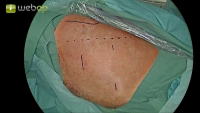
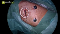
    Anatomical landmarks serve as orientation. The dashed line describes the course of the latissimus dorsi muscle. The 5th rib is usually located on an imaginary line between the scapula tip and the nipple. In the 4th intercostal space above, a 5 cm minithoracotomy is performed with the insertion of a wound protection retractor. Subsequently, under camera view, the incisions for the additional trocars can be made in the area of the 7th or 8th intercostal space ventrally and dorsally.

    Note:

    • With atraumatic technique and in skilled hands, it may also be advantageous to first make the caudal incision. After camera exploration through the caudal access, the placement of the minithoracotomy as a working access can be optimally adapted to the anatomical conditions.
  2. Preparation of the visceral pleura in the interlobium and hilum

    Video
    Preparation of the visceral pleura in the interlobium and hilum

    Repositioning the camera into the ventral incision. Initiation of preparation by incising the visceral pleura in the area of the fissure. Since the interlobar part of the pulmonary artery cannot be immediately visualized, the operation continues with the preparation of the medial lung hilum and visualization of the inferior pulmonary vein.

    Note:

    • The sequence of preparation of the pulmonary ligament, lung hilum, and fissure is variable according to the anatomical conditions and the experience of the respective surgeon.
  3. Dissection of the pulmonary ligament

    Video
    Dissection of the pulmonary ligament

    By gently pulling the lower lobe cranially, the pulmonary ligament can be stretched and transected using an ultrasonic scalpel. During this process, the lymph nodes of station 9 according to the IALSC (= International Association for the Study of Lung Cancer) can be removed. The dissection is complete when the lower pulmonary vein is exposed.

    Note:

    • Occasionally, it is necessary to press the diaphragm caudally with another swab. Care must be taken during positioning not to exert too much pressure on the abdomen, which could cause diaphragmatic elevation and difficult operating conditions.
    • After transection of the pulmonary ligament, the lower lobe is significantly more mobile, facilitating further preparation.
  4. Circular visualization of the inferior lobe vein

    Video
    Circular visualization of the inferior lobe vein

    The inferior pulmonary vein was exposed medially in the second step and caudally in the third surgical step. Now, the already partially exposed inferior pulmonary vein can be circumferentially dissected and thus prepared for later dissection. At least partial exposure of the superior pulmonary vein is essential.

    Note:

    • The sequence of preparation, whether starting with the pulmonary artery or the inferior pulmonary vein, also depends on the situs. In cases of obliterated fissure and difficulties in exposing the pulmonary artery, prior resection of the vein can also help achieve better mobility of the lower lobe and thus improve visibility.
  5. Dissection of the bridging parenchymal in the interlobium

    Video
    Dissection of the bridging parenchymal in the interlobium

    In the gap between the upper and lower pulmonary veins, the pars interlobaris of the pulmonary artery is already visible. Since the pulmonary artery was not well visualized in the interlobium, partial transection of the parenchymal bridges of the lobe fissure is performed here. Care should be taken to transect only the medial portions up to the already displayed section of the pulmonary artery.

    Note:

    • If the lobe fissure is very well developed, the preparation of the pars interlobaris of the pulmonary artery can also be performed before the parenchymal transection after preparation of the visceral pleura (Step 2).
Visualization of the pulmonary artery and the lower lobe bronchus

Through careful dissection, the parenchyma can be bluntly separated from the pulmonary artery. Subs

Activate now and continue learning straight away.

Single Access

Activation of this course for 3 days.

€7.99 inclusive VAT

Most popular offer

webop - Savings Flex

Combine our learning modules flexibly and save up to 50%.

from €3.29 / module

€39.50 / yearly payment

price overview

thoracic

Unlock all courses in this module.

€7.42 / month

€89.00 / yearly payment