
Reinforcement of the posterior wall of the inguinal canal by laparoscopic insertion of a synthetic or biological mesh placed preperitoneally.
-
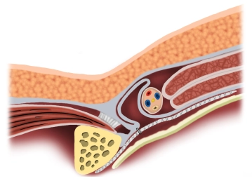
Principle
![Principle]()
-
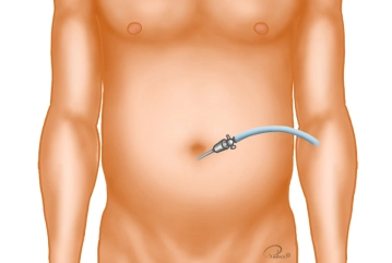
Creation of Pneumoperitoneum
![Creation of Pneumoperitoneum]()
A periumbilical skin incision approximately 1 cm long is made. Through this, the Veress needle is introduced, and the pneumoperitoneum is established. In cases of previous abdominal surgeries, the camera trocar is bluntly introduced via a mini-laparotomy.
-
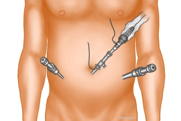
Trocar positioning
![Trocar positioning]()
The optical trocar (10 mm) is introduced bluntly with scissors after entering the abdomen and the abdominal cavity is inspected. Then, under direct vision, additional trocars are inserted laterally on both sides approximately at the level of the navel: a 5 or 10 mm trocar on the hernia side, and a 5 mm trocar on the opposite side.
Tip: The camera is guided so that the 30° optics face ventrally. This is the only way to obtain an overview of the posterior inguinal region.
Remark 1: If no inguinal hernia is visible upon inspection of the inguinal region, preparation should still be carried out, as the symptoms could be caused by the prolapse of a spermatic cord lipoma.
Remark 2: After placing the trocars, the operating table is positioned in the Trendelenburg position so that the intestines can be shifted to the upper abdomen and tilted 20° towards the surgeon to allow for better ergonomic working conditions.
Peritoneal incision and preparation, presentation of the anatomical "landmarks"
The incision of the peritoneum begins after palpation from the outside in the area of the anterior
The incision of the peritoneum begins after palpation from the outside in the area of the anterior
Activate now and continue learning straight away.
Single Access
Activation of this course for 3 days.
€7.99 inclusive VAT
Most popular offer
webop - Savings Flex
Combine our learning modules flexibly and save up to 50%.
from €3.70 / module
€44.50 / yearly payment
general and visceral surgery
Unlock all courses in this module.
€12.42
/ month
€149.00 / yearly payment