Start your free 3-day trial — no credit card required, full access included

Ileostomy placement

  1. Stoma Marking

    Video
    Stoma Marking
    Soundsettings

    Ideally, the marking and subsequent instruction for stoma care should be performed by specially trained stoma therapists or an experienced surgeon.

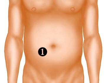
    1. Trial marking on the patient who is lying down or already seated in the area of the right rectus abdominis muscle (navel height) in a 10 × 10 cm skin area, preferably without folds, scars, or bony protrusions.
    2. Verification of the intended position in motion (standing, bending).
    3. The chosen site should be easily visible and accessible to the patient and should be compatible with the position of the pants or belt.
    4. Determining an alternative marking is recommended in case of intraoperative complications.
    5. Covering the marking with a skin-friendly adhesive tape.

    The position of the stoma significantly influences handling and care, and thus the patient's quality of life!

  2. Locating the Terminal Ileal Loop

    Locating the Terminal Ileal Loop
    Soundsettings

    After opening the abdominal cavity or upon completion of the primary operation, the terminal ileal loop is located for the creation of a loop ileostomy. The loop selected for stoma creation should be at least 20 cm away from the ileocecal valve.

  3. Suspension of the Ileal Loop

    Suspension of the Ileal Loop
    Soundsettings

    In the selected loop, an incision of the mesentery is made near the intestine, preferably in a vessel-free area, and a silicone tube is passed through.

  4. Skin Incision for Stoma and Preparation to the Fascia

    Skin Incision for Stoma and Preparation to the Fascia
    Soundsettings

    In the area of the preoperatively marked region, the circular skin incision is made after grasping the skin with, for example, a Backhaus clamp. The skin incision should not be larger than 2-2.5 cm. Subsequently, preparation to the fascia is performed.

    Note: In laparoscopic stoma creation, it is advisable to maintain the pneumoperitoneum during the formation of the stoma channel. This makes the abdominal wall thinner and facilitates the identification of each subsequent layer. Additionally, the risk of accidental injury is reduced.

  5. Splitting the Anterior Layer of the Rectus Sheath

    Splitting the Anterior Layer of the Rectus Sheath
    Soundsettings

    After excision of the skin and subcutaneous tissue, the anterior layer of the rectus sheath is split in a cruciform manner. The fascia is tensioned to avoid a curtain phenomenon.

    Note: The removal of subcutaneous tissue is not generally recommended in the literature. There is concern about a higher rate of stoma retractions due to increased subcutaneous scarring and an aesthetically unfavorable indentation of the scar after stoma relocation. Alternatively, the subcutaneous fat tissue is incised only in the longitudinal direction and retracted with hooks.

Separation of the Muscle

The rectus muscle is separated so that the posterior layer of the rectus sheath is exposed.Note: Th

Activate now and continue learning straight away.

Single Access

Activation of this course for 3 days.

US$9.30  inclusive VAT

Most popular offer

webop - Savings Flex

Combine our learning modules flexibly and save up to 50%.

from US$7.19 / module

US$86.28/ yearly payment

price overview

general and visceral surgery

Unlock all courses in this module.

US$14.38 / month

US$172.60 / yearly payment