Tube thoracostomy, open

Reading time readingtime 12:26 min.
  1. Skin incision

    Video
    Skin incision

    Note: For teaching purposes, the video was recorded with the patient in the lateral recumbent position.

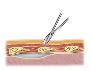
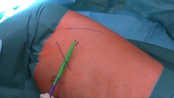
    Make a 1- to 2 cm skin incision directly at the level of the rib - in the “safe triangle” between the lower aspect of the pectoralis major muscle, the anterior aspect of the latissimus dorsi muscle, and the mammillary line.

    Tip: To avoid subcutaneous emphysema, make the skin incision just wide enough for the index finger which then can bluntly dissect the tissue and guide the tube into place.

  2. Blunt dissection

    Video
    Blunt dissection

    After the skin incision, first bluntly dissect down to the rib using the scissors. Then continue dissecting  craniad at the upper aspect of the rib.

    Tip: Tunneling through the soft tissues results in sliding layers thereby fashioning an airtight tunnel.

  3. Mini-thoracotomy

    Video
    Mini-thoracotomy

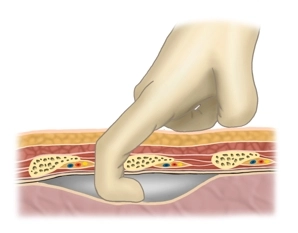
    After dissecting the muscles with the scissors strictly along the superior aspect of the rib, the parietal pleura is reached. Bluntly open it either with the finger (as shown in the illustration) or tip of the scissors. If pleural attachments/adhesions are expected palpate the pleural space with the finger. 

    Note: In patients with pneumothorax, the escaping air will make a “hissing” sound at this point; in patients with a hemothorax or pleural effusion, the fluid will drain.

  4. Inserting and positioning the tube

    Video
    Inserting and positioning the tube

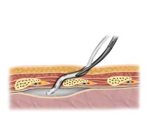
    Without its stylet grasp the tube with dressings forceps  and advance it approximately 20 cm into the chest cavity, possibly guiding it with the index finger (craniad in pneumothorax, posteroinferiorly in pleural effusion).

    Note: Leakage of fluid (hemothorax or pleural effusion) or tube condensation (pneumothorax) signals correct positioning of the tube. 

  5. Anchoring suture

    Video
    Anchoring suture

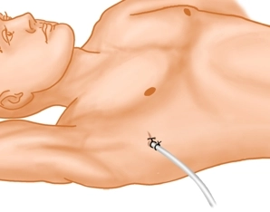
    U-suture for leakproof wound closure and fixation, connection with the suction system; radiographic monitoring.